News

... the more they stay the same.

The Neurotoolbox will be moving to the University of Oklahoma Health Science Center, starting March 2024. A tempory mirror lab website is here:

https://sites.google.com/view/neurotoolbox

The lab will continue to expand the toolbox for studying the brain and biology. We'll be hiring, so please contact us through the pathways on the contact page of either website.

Thanks to everyone at Duke (trainees, faculty, and students) who supported the Neurotoolbox. It's been fun and we'll cherish the memories. Looking toward the future, Warp one, engage!

We're very grateful to the NIH and NINDS for supporting our research. As a summary, we will be performing mesoscopic voltage imaging in the mammalian cortex with our fluroescent voltage sensors.

A press release is here and here.

We're also grateful to receive BRAIN initiative funding to perform large scale voltage imaging in zebrafish.

Open positions at the postdoctoral and graduate student levels are available. Please contact the lab using the information here.

Sometimes we take pictures of objects that are not neurons; they're still pretty good looking:

OD4 filter

1/4000 exposure

f/16

f = 300 mm

The neurotoolbox lab has had a busy summer. In all, we obtained several grants:

  • Beckman Foundation Young Investigator Award
  • Valle Foundation Young Investigator Award
  • Brain Research Foundation Seed Grant
  • DIBS Incubator Award

In addition, Jun Jiang received the prestigious Duke BME Second Year Fellowship.

We also welcomed a new postdoc, Diming Zhang to the lab.

With all of that work, we're poised to do new science. As always, we're actively recruiting graduate students and postdocs to join us. Please follow the directions here and contact us with the relevant information.

We have recently developed a new voltage sensor, Ace-mNeon, that is able to detect action potentials in live animals [local abstract][Full article at Science Magazine]. The field of GEVIs has been ongoing for almost two decades now, with the goal of being able to measure voltage signals in live animal models. There has been many developments within the past few years, each inching closer toward the goal of being able to detect action potentials in live model organisms. Much like our previous FRET-opsin sensors, we fused a bright fluorescent protein to a fast voltage sensing domain. This resulted in a sensor with 5-6 times faster kinetics than our previous MacQ-mCitrine sensors, and approximately twice as bright.

With the development of this sensors, we've demonstrated for the first time optical detection of spiking activity with a GEVI in an awake, behaving mice and flies (Fig. 1). In addition, we demonstrated that we can use this sensor to examine the propagation of voltage in sub-cellular compartments as well (Fig. 2). Although we can only do this in a small number of neurons simultaneously, breaking across the threshold of doing live animal experiments suggests that there are useful experiments that neuroscientists can immediately attempt in the near future.  Our development also suggests that large-scale parallel voltage imaging will be possible farther in the future.

optically detected spikes in live mice and flies in response to external stimuli

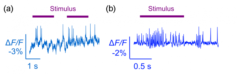
Fig 1 - (a) Optically detected spikes from an awake behaving mouse driven by visual stimulus. (b) Optically detected spikes from an awake behaving fly drive by odor stimulus.

voltage propagation through a fly neuron

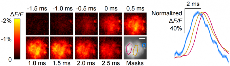
Fig 2 - Voltage propagation throughout a fly odor neuron. The voltage initially starts in the cell body (upper right of each panel), and propagates leftward through the periphery of the neuron. Masked regions at the soma (cyan) and neuron process (gold, magenta), show temporally offset voltage activity (right inset).


There will be positions open for prospective undergraduates in the fall. Students interested in the Pratt Fellows Program should contact the lab during the summer or the first week of school. It is expected that the student joins the lab for the Fall term (i.e. Fall 2015) before starting the Pratt program with the lab in the Spring term (i.e. Spring 2016). Attempting the course below will greatly help in getting up to speed for research in the lab. Projects in Protein engineering, Optical Microscopy, and advanced Image Processing are available.

Yiyang will be teaching BME590-03 in Fall 2015: The Biophysics of Neuroscience Tools. This course introduces students to the various modern tools used to study the function of the brain, and the underlying biophysics of these tools. Content will focus on novel technologies and techniques that employ electrophysiology and optogenetics, and specific topics include: patch-clamp electrophysiology, multi-electrode recordings, fluorescent proteins, optically excitatory or inhibitory rhodopsins, optical microscopy, fluorescent protein sensors, genetic delivery technologies, and numerical analysis or simulation of brain activity. The course will use lectures and recently published scientific literature to probe the capabilities and applications of these tools in current neuroscience experiments. Advanced undergraduates and enrolling graduate students interested in neuroscience and engineering should take the course.

We welcome our first batch of student researchers via the Duke Biology Department CUBR program! HalaClaireHenry, and David join us from varied backgrounds. They will attempt to learn more about molecular biology and apply their coursework to wet-lab experience.

We are looking for capable students and post-doctoral scholars to join the lab. A nascent lab is a great opportunity for students to build experiments from the ground up, and to guide exciting projects with high degrees of flexibility and responsibility. Interdisciplinary projects in the brain sciences require skills from the fields of molecular biology, neuroscience, phyics, optics, and data science, so we will always seek applicants with a wide variety of skill sets to advance our research. Prospective applicants will mostly fall into the following categories:

  • Prospective graduate students to the Department of Biomedical Engineering should apply through the BME department with emphasis in Neuroengineering. Because BME directly admits students into labs, it is highly recommended to explicitly state research directions within our lab in the statement of purpose and in the research interest sections of the application.
  • Graduate students currently at Duke University in the Departments of Biomedical Engineering, Electrical/Computer Engineering, or Neurobiology seeking a rotation should email the lab with a CV or a summary of recent research experience.
  • Prospective postdoctoral scholars should email the lab with a CV and a statement of research interests and how those interests fit with the lab direction.
  • Undergraduates currently at Duke University or from outside schools should email the lab with a statement of research interest that includes relevant coursework and future research plans. Applicants to the Pratt Research Program or REU should outline a research timeline in the inquiry.